 NEWS
NEWS
Over-expression of a protein responsible for neuronal damage in Down’s Syndrome sufferers
Press Release
- The study coordinated by the Centre for Genomic Regulation (CRG) reproduced the same morphological and functional patterns of neuronal connections in a transgenic mouse as seen in people with Down’s syndrome.
- Regulating the activity of this protein produced very similar neuronal growth to that in a healthy mouse.
The neural dendritic spine structures make connections between neurones possible (the neuronal synapses). In the case of patients with Down’s syndrome, the morphology of the neuronal information-receiving system, the dendritic tree, is altered. The dendrites are shorter and the trees are less complex, reducing and altering the flow of information via the neurone endings. It is possibly this that inhibits the development of certain normal cognitive skills, one of the characteristics of people with Down’s syndrome.
Three copies of the DYRK1A gene located on chromosome 21 are present in Down’s syndrome sufferers. In the study published in the journal Cerebral Cortex, the researchers found that the over-expression caused by excess copies of this protein in mice produces a dendritic morphology similar to that seen in the brains of people with Down’s syndrome. In these mice, the dendritic spines displayed an immature morphological aspect impeding the normal development of synaptic connections. Additionally, the activity of these neurones was reduced. This would not only directly affect the processing of information in the brain, the so-called computing power of the brain, but also the neuronal plasticity, reducing the capacity for learning.
"We see that in transgenic mice with excess quantities of this protein, the dendritic branching process in newborns alters in a very similar way to that displayed by a human with Down’s syndrome" says Mara Dierssen, head of the Neurobehavioral Phenotyping of Mouse Models of Disease group at the CRG. "This discovery may help scientists find new molecular therapeutic targets to aid not only the treatment of Down’s syndrome but also other pathologies involving intellectual disability, such as Fragile X syndrome.
The research was conducted in collaboration with various institutions, including the Cajal Laboratory of Cortical Circuits of the Technical University of Madrid, CIBERER, CIBERNED, the Faculty of Medicine at the University of Barcelona and the Laboratory of Developmental Psychology at the University of Amsterdam with the aid of the Ministry of Science and Innovation, the Ministry of Health and the European "CureFXS" project.
Reference: Martínez de Lagrán et al. Dyrk1A Influences Neuronal Morphogenesis Through Regulation of Cytoskeletal Dynamics in Mammalian Cortical Neurons. Cerebral Cortex (2012) doi:10.1093/cercor/bhr362
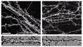
Image available at: ftp://jsarasua:jsa32rasua@perelman.crg.es Name: fig_cerebcortex_Medio.jpg
Photo footer: In culture, the neurones which over-express Dyrk1A display reduced dendritic spine density (right panel). In the lower image it can be seen that they are finer and more elongated, as in the brains of people with DS, which affects information processing.
For further information: Juan Sarasua, Press Office, Communication and PR Dept., Centre for Genomic Regulation (CRG), Tel. +34 93 316 02 37, email: juan.sarasua@crg.eu