 Decoding chromatin and DNA structure in cells undergoing reprogramming and differentiation, using super-resolution microscopy
Using super-resolution fluorescence microscopy (stochastic optical reconstruction microscopy; STORM) (Rust et al., Nature Methods, 2006) in collaboration with the group of Melike Lakadamyali (Institute of Photonic Sciences, Barcelona) we have dissected out the nanoscale organization of nucleosome assembly, with high molecular specificity and spatial resolution in a variety of somatic and stem/ reprogrammed cells. We discovered that nucleosomes are arranged into discrete groups, which we called ‘nucleosome clutches’ (as an analogy with egg clutches). While somatic cells have dense and compacted clutches that contain tens of nucleosomes, clutches in pluripotent cells contain fewer and less compacted nucleosomes. Our findings have delineated a novel model of chromatin fiber assembly, and the relationship among the decoded structure and naïve pluripotency (Ricci et al. Cell 2015).
We are currently studying the changes in chromatin structure and organization during somatic cell reprogramming and differentiation, to determine how chromatin fibers can be rearranged to overcome epigenetic barriers to gain pluripotency.
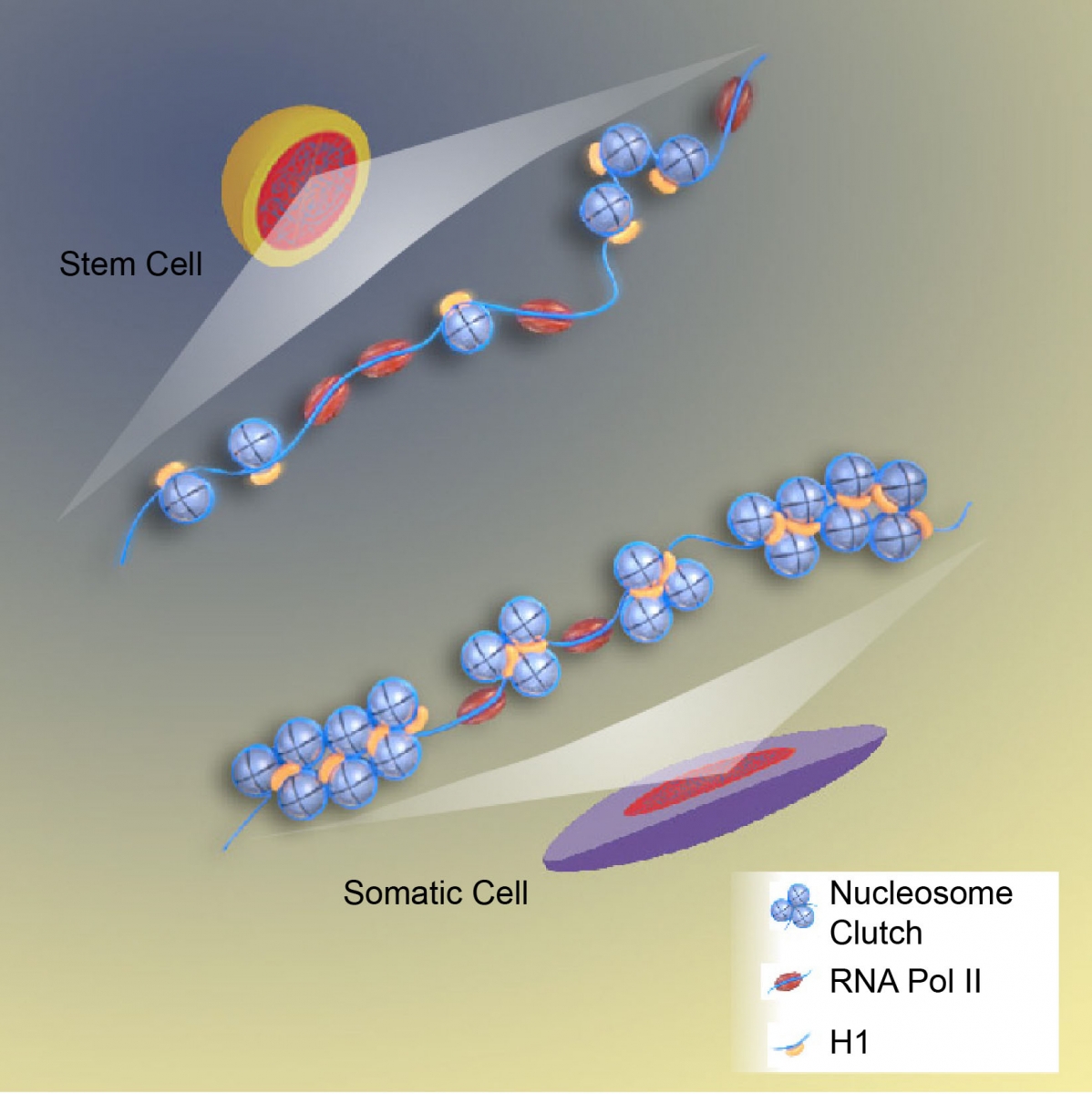
Figure above: Super Resolution Imaging reveals a new structure of chromatin fibers. Taken from Ricci et al. Cell 2015